
Valve Anatomy & Function
What are heart valves?
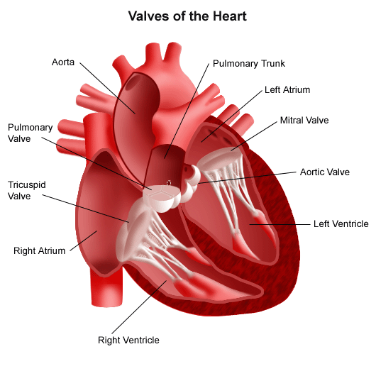
The heart consists of four chambers, two atria (upper collecting chambers) and two ventricles (lower pumping chambers). There is a valve through which blood passes before leaving each chamber of the heart. The valves prevent the backward flow of blood. These valves are actual flaps that are located on each end of the two ventricles (pumping chambers of the heart). They act as one-way inlets of blood on one side of the ventricle and one-way outlets of blood on the other side of the ventricle.
There are four heart valves:
- Tricuspid valve: located between the right atrium and the right ventricle
- Pulmonary valve: located between the right ventricle and the pulmonary artery
- Mitral valve: located between the left atrium and the left ventricle
- Aortic valve: located between the left ventricle and the aorta
How do heart valves function?
As the heart muscle contracts and relaxes, the valves open and close, letting blood flow into the ventricles and atria at alternate times. The following is a step-by-step illustration of how the valves function normally in the left ventricle:
As the left ventricle contracts, the aortic valve closes and the mitral valve opens, to allow blood to flow from the left atrium into the left ventricle.
Initially, unoxygenated blood from the body collects in the right atrium. It then flows to the right ventricle through the tricuspid valve. As the right ventricle begins to contract, the tricuspid valve closes and the pulmonary valve opens, allowing blood to flow to the lungs where it is oxygenated. After flowing through the lungs, this newly oxygenated blood collects in the left atrium.
As the left atrium contracts, blood flows into the left ventricle. When the left ventricle contracts again, the mitral valve closes and the aortic valve opens, so blood flows into the aorta to travel to the body.
Click to view larger